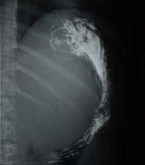
Giant cell tumour in lateral condyle of femur.
A 31 years old female presenting with knee pain since last 6 months with history of fall and inability to walk for 2 days from the time of examination.
CT scan and MRI were done simultaneously, whihc reveal an eccentric, expansile lytic lesion with corticla thinning and a pathological fracture in laterla condyle of femur with joint effusion. Here are the images. Final diagnosis is Giant cell tumour of lower end of femur.
Giant cell tumous are usually seen after the closure of growth plates and are seen between the age of 20-50 years with peak incidence between 20-30 years with a femle predeliction. They typically arise from the metaphysis of long bones, extend into the epiphysis adjacent to the joint surface, and have a narrow zone of transition.


Comments
Post a Comment